Parvovirus B19 is a single stranded DNA virus, which is transmitted by respiratory droplets or blood.
The virus produces a mild, self-limiting infection in adults, but can cause spontaneous miscarriage or intrauterine death if transmitted to the fetus during pregnancy.
Approximately 1 in 400 women become infected with parvovirus B19 during their pregnancy, with vertical transmission to the fetus in up to 33% of cases. Spontaneous miscarriage or intrauterine death occurs in approximately 9% of infected pregnancies.
In this article, we shall look at the clinical features, investigations and management of parvovirus B19.
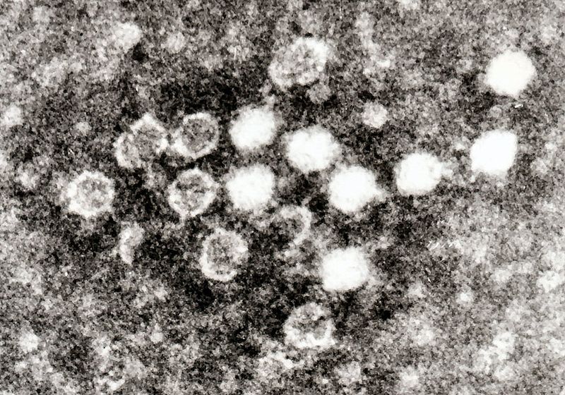
Fig 1 – Electron micrograph of negatively stained Parvovirus B19 in blood. Each virus particle is about 25 to 30nM in diameter.
Clinical Features
Parvovirus infection in adults is usually asymptomatic. The most common clinical feature is symmetrical arthralgia – typically of the proximal interphalangeal joints and/or knees.
In children, parvovirus infection manifests with:
- Upper respiratory tract infection
- Malaise
- Headaches
- Low-grade fever.
- Erythema infectiosum (slapped cheek syndrome) – a maculopapular rash sparing the nose, eyes and mouth.
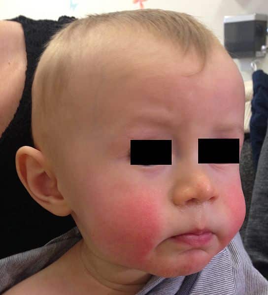
Fig 2 – Slapped cheek syndrome, seen in parvovirus B19 infection.
Investigations
In cases where a mother has potentially come into contact with parvovirus B19, viral serology can be performed:
- Parvovirus specific IgM antibodies indicate recent infection
- Parvovirus specific IgG antibodies indicate past infection and therefore immunity.
Management
Women with a confirmed infection of parvovirus B19 should be referred to a fetal medicine specialist for further management.
Maternal
Parvovirus infection is self-limiting, and does not require treatment. Antipyretics and analgesia can be given.
Fetal
The main risk of fetal parvovirus infection is fetal hydrops – the abnormal accumulation of fluid in two or more fetal compartments.
Management is typically:
- Serial ultrasound scans and Doppler assessment
- Starting 4 weeks post infection or at 16 weeks.
- Repeated every 1-2 weeks, until 30 weeks gestation.
- If there is evidence of fetal hydrops on ultrasound, the patient should be referred to tertiary centre for intrauterine erythrocyte transfusion.
Fetal Hydrops
Fetal hydrops is the most common manifestation of fetal parvovirus B19 infection.
It is diagnosed by ultrasound scan with features including: ascites, subcutaneous oedema, pleural effusion, pericardial effusion, scalp oedema and polyhydramnios.
Fetal hydrops occurs because parvovirus B19 has an affinity for the erythroid system and replicates within the erythroid progenitor cells of the liver and bone marrow. This induces severe anaemia which results in:
- High output cardiac failure
- Increased extrahepatic and hepatic erythropoiesis – resulting in portal hypertension and hypoproteinaemia with subsequent ascites.