Oligohydramnios refers to a low level of amniotic fluid during pregnancy.
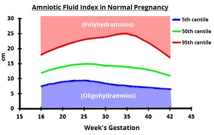
It is defined by an amniotic fluid index that is below the 5th centile for the gestational age, and is thought to affect approximately 4.5% of term pregnancies [AJOC, 2004].
In this article, we shall look at the aetiology, investigations and management of oligohydramnios.

Fig 1 – Amniotic fluid centiles during pregnancy. Polyhydramnios is over the 95th centile, oligohydramnios is below the 5th centile
Pathophysiology
The volume of amniotic fluid increases steadily until 33 weeks of gestation. It plateaus from 33-38 weeks, and then declines – with the volume of amniotic fluid at term approximately 500ml.
It is predominantly comprised of the fetal urine output, with small contributions from the placenta and some fetal secretions (e.g. respiratory).
The fetus breathes and swallows the amniotic fluid. It gets processed, fills the bladder and is voided, and the cycle repeats. Problems with any of the structures in this pathway can lead to either too much or too little fluid.
Anything that reduces the production of urine, blocks output from the fetus, or a rupture of the membranes (allowing amniotic fluid to leak) can lead to oligohydramnios.
Aetiology
The main causes of oligohydramnios are:
- Preterm prelabour rupture of membranes
- Placental insufficiency – resulting in the blood flow being redistributed to the fetal brain rather than the abdomen and kidneys. This causes poor urine output.
- Renal agenesis (known as Potter’s syndrome)
- Non-functioning fetal kidneys, e.g. bilateral multicystic dysplastic kidneys
- Obstructive uropathy
- Genetic/chromosomal anomalies
- Viral infections (although may also cause polyhydramnios)
Diagnosis of Oligohydramnios
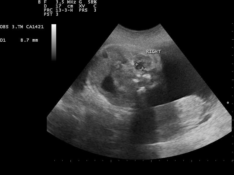
The diagnosis of oligohydramnios is made via ultrasound examination. There are two ways of measuring amniotic fluid; amniotic fluid index (AFI) or maximum pool depth (MPD). They have similar diagnostic accuracy, however AFI is more commonly used.
- Amniotic fluid index is calculated by measuring maximum cord-free vertical pocket of fluid in four quadrants of the uterus and adding them together.
- Maximum pool depth is the vertical measurement in any area.
Clinical Assessment
Oligohydramnios is a diagnosis made via ultrasound examination. Therefore, the clinical assessment of the patient is directed at establishing any underlying cause:
- History
- Inquire about symptoms of leaking fluid and feeling damp all the time (often described as new urinary incontinence).
- Examination
- Measure the symphysis fundal height.
- Perform a speculum examination (can a ‘pool’ of liquor be seen in the vagina?).
- Ultrasound
- Assess for liquor volume, structural abnormalities, renal agenesis and obstructive uropathy.
- Measure fetal size. Small babies can result from placental insufficiency, which also causes oligohydramnios. There may also be a rise in pulsatility index of the umbilical artery Doppler in placental insufficiency.
- Karyotyping (if appropriate) – particularly in cases of early and unexplained oligohydramnios.
When considering ruptured membranes as a cause for oligohydramnios, a bedside test can be performed to detect the presence of IGFBP-1 (insulin-like growth factor binding protein-1) or PAMG-1 (placental alpha-microglobulin-1) in the vagina. These proteins are found in amniotic fluid, and if detected, strongly suggest membrane rupture.
Management
The management of oligohydramnios is largely dependent on the underlying cause. The two most common causes are rupture of the membranes and placental insufficiency.
Ruptured Membranes
If oligohydramnios is due to ruptured membranes, labour is likely to commence within 24-48 hours in most pregnancies.
In cases of preterm rupture of membranes (i.e. before 37 weeks’ gestation), and where labour doesn’t start automatically, induction of labour should be considered around 34-36 weeks (in the absence of infection).
A course of steroids should be given to aid fetal lung development, and antibiotics to reduce the risk of ascending infection.
Placental Insufficiency
In women where oligohydramnios is caused by placental insufficiency, the timing of delivery depends on a number of factors:
- Rate of fetal growth
- Umbilical artery and middle cerebral artery Doppler scans
- Cardiotocography
These babies are likely to be delivered before 36-37 weeks.
Prognosis
Oligohydramnios in the second trimester carries a poor prognosis. In the majority of these cases, there is premature rupture of membranes (which may or may not be associated with infection), with subsequent premature delivery and pulmonary hypoplasia – which can cause significant respiratory distress at birth
When oligohydramnios is associated with placental insufficiency, there is also a higher rate of preterm deliveries (usually through planned induction of labour). These cases will carry a poorer prognosis than that of a normally grown fetus.
Amniotic fluid also allows the fetus move its limbs in utero (exercise). Without this, the fetus can develop severe muscle contractures – which may lead to disability despite physiotherapy after birth.
Summary
- Oligohydramnios occurs when the amniotic fluid is < 5th centile for gestational age.
- The most common causes are premature rupture of membranes (often missed by the mother) and placental insufficiency, however structural abnormalities such as renal agenesis should be considered.
- Prognosis is linked to gestation at diagnosis and likely development of pulmonary hypoplasia and premature delivery.
- Treatment is by optimising gestation of delivery.

